Meet Armin Khamoshi
|
Major: Physics and Pure Mathematics
Graduation Year: December 2016 Field of Study: Mathematical Modeling and Numerical Simulation (Advanced Imaging department) Abstract: About MR Spectroscopy: Medical imaging techniques, in general, are designed to provide information from internal aspects of the body. Along with many of these techniques, magnetic resonance imaging (MRI) uses high magnetic field to image different anatomical structures or pathologies. In-vivo Magnetic Resonance Spectroscopy (MRS) is a specialized technique associated with MRI with emphasis on tracking metabolic changes and reaction rates (kinetics) in the brain, liver, heart and many other organs. Studying Kinetics of Metabolites Using MRS Data: The spectra acquired by MR spectroscopy ordinarily feature a number of Lorentzian or Gaussian lineshapes (symmetric "bell curve" peaks) with some random noise near the base-line (e.g. Figure-1). Each peak corresponds to a certain chemical nuclei whose nuclear magnetization resonates at that frequency. The area under the graph of each peak (peak intensity) is proportional to the concentration of that molecular specie. When studying metabolism of a certain organ is concerned, biologists can use this phenomenon to investigate the kinetics of each molecule by tracking the time evolution of the intensities of the chemicals through several data acquisition over a period of time. The information gathered in this process sheds light on how an organ consumes some labeled substrates under different pathological conditions. |
|
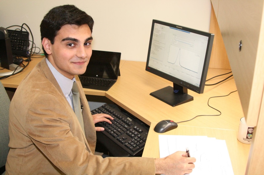
Using Computer-Programming to Calculate the Kinetics: While finding the kinetics of certain metabolites are motivated from the biomedical perspective, it is a cumbersome process to calculate the kinetics of each chemical in a non-automated fashion—namely correcting the phase, manual peak identification, specifying the boundary, manual peak-fitting, problem of convergence etc. The issue is even worse for in-vivo experiments; for 1) the signal to noise ratio is comparably lower 2) the spectrum cannot always be phase corrected. Therefore, we aimed to solve the aforementioned difficulties by implement computer programs that requires the least number of manual interventions for data processing.
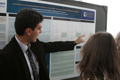
In this communication: We demonstrate our approach for automating the calculation of kinetics by evaluating its performance on the data from metabolism of hyperpolarized DHA in Rat liver. We used several functions and toolboxes provided in MATLAB to fit our model to data, and to integrate the desired boundaries and find the kinetics. The theory underlying the model and architecture of our code is also explained here. Our ultimate goal is to create a toolbox which, given any set of MR spectra, reads the data, identifies the peaks, calculates the kinetics and plots the result. |
My Experience:
A semester long full-time exposure to research was certainly more insightful than solely gaining proficiency in some scientific endeavors. It has given me, and certainly other fellows, a more realistic understanding of the scientific research environment. My personal experience was absolutely delightful. My project incorporate my passion in mathematics, physics, and computer science, and I enjoyed every second of working on it. Had it not been provided by the Green Fellowship, I probably would not have had such a productive and insightful experience. |
My Advice:
I want to use this opportunity to encourage students in physics and mathematical sciences to participate in this program. There are plenty of opportunities, and also demand, for students with background in mathematics and computer programming. |
